"Volumetric AI-driven measurement clearly represents the future of aneurysm management. The paper highlights the power of this extraordinary tool and what it might mean for patient care." -- Dr. Daniel Sahlein
In their retrospective study, Dr. Daniel Sahlein and co-authors found that the AI-enabled aneurysm measurement tool, Rapid Surgical Preview, had a higher sensitivity for detecting changes in aneurysm size than the current clinical practice (manual linear measurement).
In current clinical practice, the maximum diameter of the aneurysm is used to determine aneurysm size. However, aneurysm morphology is dynamic. Manual linear measurements do not accurately capture the size of an aneurysm.
The AI-enabled aneurysm measurement tool, Rapid Surgical Preview, calculates reproducible volumetric and surface area measurements, which is a more accurate reflection of aneurysm size and hence aneurysm growth. It represents a revolution in the technique for measuring aneurysm size and helping physicians assess rupture risk with proper surveillance and quantification of aneurysms.
Growth detected in aneurysms using manual linear measurement
From a single-practice database of over 5000 intracranial aneurysm patients, five patients who met the inclusion criteria were selected for the study. These patients had at least two cross-sectional neurovascular imaging studies before experiencing the aneurysm rupture. The average time between the two imaging studies was 3.71 years. The aneurysms ruptured during conservative management.
The mean age of the patients was 62.2 years, and they had variable medical histories at the time of imaging. Four patients had saccular aneurysms, and one patient had a fusiform aneurysm.
Growth was recorded in three out of five aneurysms.
AI-enabled measurement tool identified growth in volume, surface area, and maximum size of aneurysms
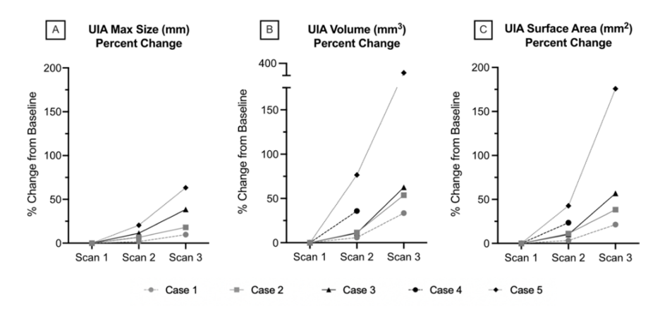
In contrast to the current clinical practice, the AI-enabled Rapid Surgical Preview identified the following:
 Figure 1. Two sets of scans from five patients who had unruptured intracranial aneurysms were processed using Rapid Surgical Preview to measure the changes in maximum dimension, volume, and surface area over time. Chart source: http://dx.doi.org/10.1136/jnis-2022-019339
As the chart above shows, the minimum relative increase in the maximum dimension was close to 1.8%. In contrast, the minimum relative increase in volume was almost 6 %, underscoring the limitations of using the maximum linear dimension to determine aneurysm growth.
Profound implications for aneurysm management
The results of this study open the possibility that all or the vast majority of aneurysms that rupture could be enlarging — a finding with far-reaching implications. Additional studies should be done to further explore the use of AI tools like Rapid Surgical Preview to help physicians better assess rupture risk with proper surveillance and quantification of aneurysms. RapidAI is encouraged to see and participate in these types of ground-breaking studies.
Learn more about the Rapid Surgical Preview, an FDA-cleared imaging platform for comprehensive cerebral aneurysm management. Read how clinical leaders in the field are reacting to this ground-breaking study here.


