MRI Mismatch
The mismatch map provides a comparison between brain regions with substantial reductions in tissue diffusion in pink and regions with significant hypoperfusion in green as reflected by delays in contrast arrival time (Tmax delays).
Rapid MRI automatically delivers clinically-proven, advanced MR diffusion and perfusion image analysis to help in the assessment of salvageable brain tissue.
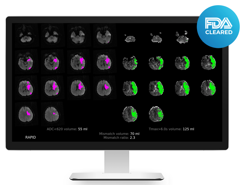
Rapid MRI automatically delivers quantified, color-coded MR diffusion and perfusion maps to help stroke teams quickly assess patients who may benefit from endovascular thrombectomy.
The mismatch map provides a comparison between brain regions with substantial reductions in tissue diffusion in pink and regions with significant hypoperfusion in green as reflected by delays in contrast arrival time (Tmax delays).
The mismatch map can also be configured to show if a patient meets imaging criteria for mechanical thrombectomy (MT).
This map shows the severity of the delays in contrast arrival times (Tmax), the volume of tissue with delays, and the Hypoperfusion Intensity Ratio (HIR) which is automatically calculated.
It offers a co-registered view of the diffusion-weighted images with the MR perfusion maps. No thresholds are applied and no volumes are calculated on this map. It allows a comparison between DWI and perfusion images.
The AIF plot displays the location automatically chosen by the software for the arterial input function (AIF) and the venous output function (VOF). The movement plots document how much patient movement occurred during the scan.
Rapid MRI has been clinically proven to accurately identify brain regions with reduced ADC and transit time in multiple clinical trials including EXTEND, SWIFT-PRIME, and DEFUSE 3.
Within minutes of receipt of a scan, this MRI analysis software automatically delivers quantified, color-coded MR diffusion and perfusion maps that identify brain regions with reduced apparent diffusion coefficient (ADC) and transit time that exceed pre-specified thresholds correlating to ischemic core and critically hypoperfused tissue.
Rapid MRI immediately delivers advanced imaging results via PACS, email, and the Rapid mobile and web apps—helping stroke teams make faster triage-or-transfer decisions and save more lives.


"Our research at UCLA shows that in a typical stoke where there is a large vessel occlusion, two million brain cells die for every minute that goes by without treatment, underscoring just how critical a rapid response to stroke is. We are looking forward to bringing Rapid into our Mobile Stroke Unit—the first of its kind in the Western U.S. This added level of imaging analysis will lead to more efficient diagnosis and triage of patients requiring routing to advanced stroke centers directly from the field."

"Diagnosis of acute stroke is a high pressure, time-sensitive situation. The combination of the Rapid and Join platforms is a powerful tool to enable our team to confidently get the stroke patient to the right clinical treatment in this life-threatening environment."

